From Chronic to Acute: Strategies for Preventing Wound Chronicity
February 27, 2020
Wound chronicity is defined as any wound that is physiologically impaired due to a disruption in the wound healing cascade: 1) hemostasis, 2) inflammation, 3) proliferation, and 4) maturation/remodeling. To effectively manage chronic wounds, we must understand the normal healing process and wound bed preparation (WBP). Wound chronicity can occur due to impaired angiogenesis, innervation, or cellular migration. The presence of biofilm and infection are the most common causes of delayed healing.1
Biofilm extensively contributes to evolving wounds into a state of prolonged inflammation by stimulation of nitric oxide, inflammatory cytokines, and free radicals.2 Biofilms harbor physical and metabolic defenses. There’s no specific number of days that differentiates an acute wound from chronic, but some suggest the lack of approximately 15% reduction weekly or approximately 50% reduction of the surface area of the wound over a one-month period validates wound chronicity.3
Common Chronic Wounds
Determining if a patient’s wound is likely to become chronic involves performing a thorough assessment of the patient. Patients with multiple comorbidities—especially conditions such as diabetes or obesity—are more likely to experience delayed healing, as are those on certain medications (immunosuppressants) or who smoke.4 Certain wounds are more likely to become chronic than others; clinicians should learn to recognize these wounds and take appropriate steps to prevent chronicity.
- Venous ulcers – Venous ulcers develop when the valves in the veins do not properly work and allow pooling in the veins, which in turn leads to swelling and ulcer development.5
- Arterial ulcers – Arterial ulcers are caused by poor profusion of nutrient-rich blood to the lower extremities, leading the oxygen-deprived tissue to become damaged. These wounds are typically full-thickness.6
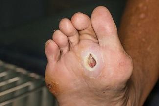
- Diabetic foot ulcers – Diabetic foot ulcers are the result of repetitive trauma to the foot, usually over areas of deformity or in those with neuropathy, limiting sensation in the foot.7
- Pressure injuries – Pressure injuries develop over bony prominences after exposure to prolonged pressure, shear, or friction.5
A holistic approach is best at assessing a patient with wound chronicity. Performing a thorough history and physical examination, wound assessment, nutritional status, self-care status, and education are key components in a comprehensive treatment plan. Identifying the correct wound etiology is paramount, but not always possible. Chronic wound types can include but are in no way limited to those listed above. Surgical or traumatic wounds, radiation injuries and malignancy, as well as others, all face the risk of becoming chronic, under the right conditions.8 Comorbidities such as diabetes, autoimmune disease, peripheral arterial disease, obesity, medications and anatomical location are all factors for higher risk in wound chronicity and infection.
Standard of Care
What’s considered standard of care may vary based on the wound’s etiology, but all wounds should generally be debrided, if necessary, monitored for infection, off-loaded as needed, and have a moist wound bed.
Wounds should be evaluated for the presence of non-viable tissue and then an appropriate debridement method chosen. Sharp/surgical debridement is often referred to as the gold standard, but depending on the patient, the level of non-viable tissue, and the goals for the wound, other methods may be preferred. Non-viable tissue inhibits the growth of healthy tissue and can also increase the risk of wound infection. Infection is the most common preventable challenge in the wound healing continuum. Clinicians must differentiate between systemic infection and spreading infection, and distinguish between incidental positive cultures and a true pathogen affecting wound healing. Bacteria is a normal part of the skin flora; however, wounds with a critical threshold of 105 bacteria per gram of tissue, has been proposed as the characterization between spreading infection and systemic infection.9,10 Wound surface cultures do not confirm or rule out continued infection; nonetheless, clinically diagnosing an infected wound is vital.10 Tissue biopsies have finer sensitivity and specificity in isolating a causative organism and are generally considered the gold standard.
Advanced wound care dressings can help to serve as a protective barrier from infection, provide autolytic debridement, and manage bacteria, thus promoting wound healing progress. Choose appropriate advanced wound care dressings considering such factors as wound depth, exudate amount, and anatomical location to maximize wound healing outcomes. There are various types of wound dressings with integrated antimicrobial compounds available to eradicate biofilm, manage bacteria, and serve as wound bed preparation.
Advanced Wound Care Options in Chronic Wounds
Choose treatments that are appropriate for the wound while using a patient-centered approach. Treatment selection can be confusing due to the wide range of products available. Always evaluate wound depth, exudate amount, wound size, wound location, and treatment availability, including payor source. Advanced wound care treatments come in various forms and sizes, so health care professionals should take the extra time to select the most appropriate dressing. Goals for wound treatment selection are bacterial balance and an optimal, moist wound healing environment. Some treatment options include the following:
- Transparent films
- Hydrocolloids
- Hydrogels
- Alginates/hydrofibers
- Collagen
- Foams/super absorbents
- Negative pressure wound therapy
- Skin substitutes – cellular and tissue-based products
- Growth factors
Health care professionals should take care to familiarize themselves with the above treatments, as each type has different properties and indications.
Conclusion
Chronic wounds are a burden and challenge to the health care system, health care professionals and patients. Implementing an early, multifactorial treatment plan can improve wound healing outcomes. Health care professionals should identify potential risk factors and help their patients manage contributing factors to chronic wounds. Utilizing various wound care dressing technologies can help chronic wounds move toward reepithelialization.
Reference
1. Golinko MS, Clark S, Rennert R, et al. Wound emergencies: the importance of assessment, documentation, and early treatment using a wound electronic medical record. Ostomy Wound Manage 2009; 55:54.
2. Frykberg RG, Banks J. Challenges in the treatment of chronic wounds. Adv Wound Care. 2015;4(9):560–82.
3. Sheehan P, Jones P, Giurini JM, et al. Percent change in wound area of diabetic foot ulcers over a 4-week period is a robust predictor of complete healing in a 12-week prospective trial. Plast Reconstr Surg 2006; 117:239S.
4. Ucciolo L, Izzo V, Meloni M, et al. Non-healing foot ulcers in diabetic patients: general and local interfering conditions and management options with advanced wound dressings. J Wound Care. 2015;24(4), 35–42.
5. DeKalb Medical. Common types of chronic wounds. http://www.dekalbmedical.org/our-services/wound-care/chronic-wounds/typ…. Accessed February 21, 2020.
6. Cleveland Clinic. Lower Extremity (Leg and Foot) Ulcers. Cleveland Clinic. http://my.clevelandclinic.org/heart/disorders/vascular/legfootulcer.aspx. Published August 17, 2017. Accessed February 21, 2020.
7. Salcido R. Pressure Ulcers and Wound Care. Medscape Reference. http://emedicine.medscape.com/article/319284-overview#aw2aab6b2. Updated June 11, 2018. Accessed February 21, 2020.
8. Brem H, Balledux J, Bloom T, et al. Healing of diabetic foot ulcers and pressure ulcers with human skin equivalent: a new paradigm in wound healing. Arch Surg 2000; 135:627.
9. Qualitative bacteriology and leg ulcer healing. Trengove NJ, Stacey MC, McGechie DF, Mata S J Wound Care. 1996 Jun; 5(6):277-80.
10. Armstrong DG, Liswood PJ, Todd WF. William J. Stickel Bronze Award. Prevalence of mixed infections in the diabetic pedal wound. A retrospective review of 112 infections. J Am Podiatr Med Assoc. 1995;85(10):533–537. doi: 10.7547/87507315-85-10-533
The views and opinions expressed in this blog are solely those of the author, and do not represent the views of WoundSource, HMP Global, its affiliates, or subsidiary companies.


